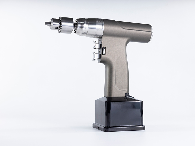
The use of medical bone drills in cardiac surgery

The usage of medical bone drills in cardiac surgery is as follows:
1. Insert left heart drainage tube: slide the suture onto the right superior pulmonary vein for purse string closure, and fix it with forceps. After the aortic occlusion tube is completely emptied and the ACT value exceeds 480s, the machine is turned around, cooled down, and the upper and lower veins are blocked. Then, bear paw forceps and aortic occlusion forceps are handed over to block the aorta. Perform coronary artery perfusion with stopping fluid, with a perfusion volume of 10-15ml/Kg. Apply ice to the surface of the heart for cooling. Cut open the heart, explore and hand over the surgical knife, hand over the cardiac forceps to cut open the heart, hand over the tissue to enlarge the incision, and hand over the atrial or ventricular hooks to appear.
2. Close the atrial incision, suture the right atrial incision continuously with a sliding thread, or suture the left atrial incision with a sliding thread, place the operating table at a low head position, and open the upper and lower veins. Open the aortic occlusion forceps, and the heart can generally resume beating automatically. In cases of difficulty in resuming beating, electric shock can be used to resume beating. After extracorporeal circulation assisted perfusion and cardiac arrest, all examination indicators were normal and stopped.
3. Parallel circulation is used to restore cardiac function. The left heart drainage tube is removed, and the left heart is suctioned and placed into an injection tube for retrograde exhaust, causing the upper chamber catheter to retract to the right atrium and restore the anal temperature to 35 degrees Celsius.
4. Extracorporeal circulation extubation: After the cardiac function returns to normal, remove the coronary perfusion tube, sequentially remove the inferior vena cava tube and the superior vena cava tube, tie the upper purse string after extubation, and if necessary, tie or suture it. Central vein infusion or use protamine and heparin at the root of the aorta. Finally, hand over the bear paw forceps, remove the aortic cannula, and connect it with a purse string.
5. Close the sternotomy caused by the medical bone drill: After inserting the chest and pericardial mediastinal drainage tubes, count the items, hand them over to the wire suture chest, and count the items again after suturing.

