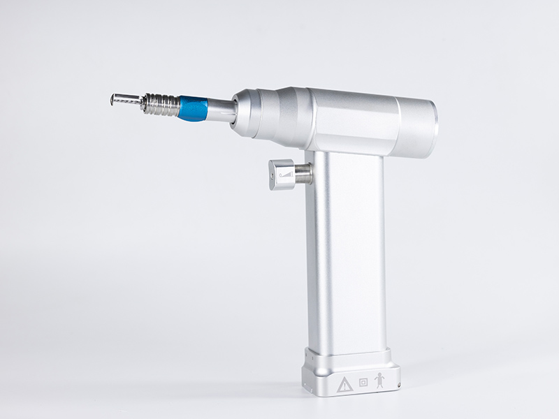
Precautions for craniotomy drill during drilling operation

The precautions for craniotomy drill during drilling operation are as follows:
Special attention should be paid to the protection of the venous sinus located above the dura mater when using a craniotomy drill. Pay attention to the integrity of the dura mater. Avoid damaging the air hole. Carefully handle lesions infiltrating the skull and bone markers hidden outside the dura mater. When using an end mill for operation, it is important to avoid transmitting the vibration of the end mill to the brain tissue (which can cause contusions and subarachnoid hemorrhage). It is also necessary to avoid forcefully opening the inner plate and inserting the drill into the dura mater and brain tissue, which may cause cortical damage.
The number and size of drill bits required for safe completion of craniotomy depend on several factors, including the selected position and size of the bone window (whether it spans the dura mater or sutures). The nature of the lesion. The age of the patient. The overall degree of adhesion of the dura mater to the inner plate of the skull. The suggestion is to perform perforation within the hairline to minimize the abnormal appearance caused by skull defects. By enlarging the perforation, the dura mater beyond the edge of the bone hole can be more effectively peeled off.
Operate within the boundary range of the collected bone window. The peeling range of the dura mater cannot exceed the edge of the bone window. Because excessive stripping of the dura mater can cause occult epidural hematoma, especially in young patients. Drill a hole along the base of the zygomatic arch. Use sufficient amount of physiological saline to eliminate obstacles on the outer plate of the skull (then carefully grind the inner plate). This will expose a portion of the dura mater. Then remove the remaining thin bone fragments from the remaining inner plate. This operation method can enlarge the inner edge of the bone hole beyond its outer edge, allowing the anterior end of the meningeal stripping ion to have the correct operating angle, which can be indirectly adhered to the inner plate to avoid dural rupture caused by misoperation.

