- Technical articles
- Current location:Home>>Technical articles
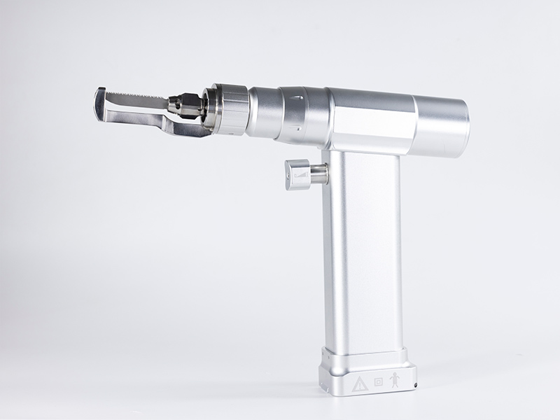
Application of sternotomy in cardiac surgery

The application of sternotomy in cardiac surgery is as follows:
(1) Cover the superior vena cava block: Hand over the cardiac forceps, cut off the endocardium of the superior vena cava and pulmonary vein fossa, separate the forceps to widen the gap, free the superior vena cava, use right angle forceps or superior vena cava free forceps to wrap around the posterior wall of the superior vena cava, and remove the block, silicone hose, and hook.
(2) Inferior vena cava occlusion band: Hand the forceps to the heart, cut off the tunica vaginalis between the right inferior pulmonary vein and inferior vena cava, and use free forceps to bypass the machine.
(3) Insert the superior vena cava tube: Hand over the cardiac forceps and traction forceps, lift the right atrial appendage, hand over the surgical knife to cut the inferior vena cava and lead out the blocking band. Thread the blocking band through the silicone tube with a silicone tube and hook, and fix it with a straight vessel clamp.
(4) Suture the superior vena cava intubation funnel: Hand over the non damaging suture, suture the superior vena cava intubation funnel on the right ear, hand over the suture hook, and fix it with mosquito forceps.
(5) Suture the funnel of inferior vena cava catheterization: suture the funnel with a non damaging suture needle in the anterior wall of the right atrium near the entrance of the inferior vena cava, pull up the hook sleeve, and secure it.
(6) After heparinization, the aorta was intubated and the adventitia of the aorta was cut at the intubation site. The cardiac forceps were used to cut off the adventitia of the aorta. The aortic cannula and surgical knife required for the transfer surgery are opened with a small opening in the pericardium, the aortic cannula is inserted, the pericardial thread is tightened, the blocking tube and aortic tube on the pericardium are fixed with a transfer rope, and after exhaust, the extracorporeal circulation is connected to the right atrial appendage or tissue. The sternotomy separates the intramural muscle trabeculae, and the appropriate superior vena cava cannula is handed over. The catheter is inserted into the superior vena cava of the right atrium from the right atrial appendage and secured with a wire.
(7) Insertion of inferior vena cava catheter: Hand over the cardiac forceps or vascular forceps to lift the right atrial wall, stretch the forceps to enlarge the incision, insert the inferior vena cava catheter, tighten the pericardial wire transfer rope, fix the pericardial wire and inferior vena cava catheter, and perform machine connection.
The above is an introduction to the application of sternotomy in cardiac surgery. Thank you for reading.

